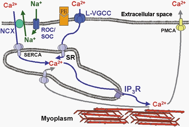
Figure 6. Model for Ca2+ movements during PE-induced [Ca2+]i oscillations in VSMCs from rabbit IVC.

IP3 generated as a consequence of α-adrenergic activation releases Ca2+ towards the myoplasm to activate the myofilaments. Subsequent Ca2+ removal occurs partially through the PMCA, while the remainder is taken up into the SR by the SERCA. ROCs/SOCs, activated by receptor activation and SR depletion, allow entry of mainly Na+ and some Ca2+. This Na+ entry raises [Na+]i in a restricted space between the plasma membrane and the SR membrane and drives NCX in the reverse mode to supply Ca2+ to SERCA to refill the SR lumen, thereby completing the cycle. PE, phenylephrine; NCX, sodium/calcium exchanger; ROC/SOC, receptor-operated channel/store-operated channel; SERCA, sarcoplasmic- endoplasmic reticulum Ca2+-ATPase; L-type VGCC, L-type voltage-gated Ca2+ channel; PMCA, plasma membrane Ca2+-ATPase; IP3R, IP3-sensitive SR Ca2+ release channel; SR, sarcoplasmic reticulum.
