
Figure 1. Properties of recombinant ρ1 receptors expressed in HEK293 cells after adeno-ρ infection.
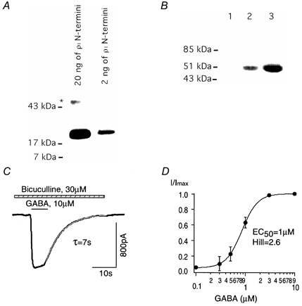
A, Western blot with N-terminal ρ1 GABAC receptor antibodies, which recognized a bacterially synthesized N-terminal fusion protein. The band indicated by the asterisk probably represents non-specific staining of an unidentified protein. B, Western blot from uninfected HEK293 cells (lane 1) and HEK293 cells infected with adeno-ρ (2 and 10 PFU cell−1, lanes 2 and 3, respectively). Note the specific band of the ρ1 subunit of the GABAC receptor (50 kDa) in lanes 2 and 3. C, whole-cell current evoked at a holding potential of −50 mV with GABA (10 μm) in the presence of bicuculline (30 μm) from HEK293 cells after adeno-ρ infection. Decay of the current upon GABA removal was well described by a single exponential component with a time constant (τ) of 7 s. D, mean dose-response relationship for GABA-activated current fitted with a Hill equation (n = 4).