
Figure 4. Recombinant ρ1 receptors expressed in cultured hippocampal slices after adeno-ρ infection.
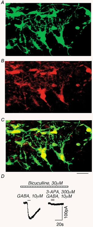
A-C, images of cultured hippocampal slices after co-infection with adeno-GFP and adeno-ρ using a confocal scanning microscope. A, GFP fluorescence. B, staining with N-terminal ρ1 GABAC receptor antibodies (red). C, co-localization of GFP and ρ1. Scale bar, 50 μm. D, example of a recombinant GABAC current in the slice after adeno-ρ infection. The current was blocked by 3-APA, an antagonist of GABAC receptors.