
Figure 2. Time- and dose-dependence of camptothecin-induced DNA fragmentation during apoptosis.
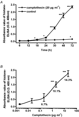
Direct cytosolic nucleosome-bound DNA was detected by histone ELISA in HT-29/B6 cells grown to confluence in 24-well dishes. Data represent means ±s.e.m. (*P < 0.05, ***P < 0.001) of 4 independent experiments (n = 4), each carried out in triplicate. A, time course of cells treated with camptothecin and controls. B, dose-response curve; cells were incubated for 48 h. Values for apoptotic nuclei (percentage of total, control = 3.5 %) were obtained by DAPI staining (n = 6).