
Figure 9. Differential effects of ω-conotoxin and nifedipine on somatostatin-induced inhibition of exocytosis.
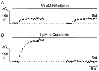
Trains of ten 500 ms voltage-clamp depolarisations from -70 to 0 mV were applied at a frequency of 1 Hz (Vm) using the perforated-patch whole-cell configuration in single rat α-cells. The experiments were performed in the presence of 10 μm forskolin and the trains of depolarisations were applied before (left) or after (right) the addition of 400 nm somatostatin (Sst) in the continuous presence of either 50 μm nifedipine (A) or 1 μmω-conotoxin (B). Note differential effects of Sst when applied in the presence of nifedipine and ω-conotoxin. The interval between the trains of depolarisations was 2 min. Data are representative of 5 separate experiments in both A and B.