
Figure 2. EPSPs evoked by dorsal column and corticofugal stimulation.
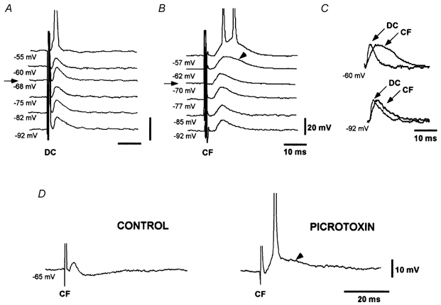
A, dorsal column EPSP (DC) evoked during depolarising and hyperpolarising current pulses show the expected decrease and increase in EPSP amplitude with depolarisation and hyperpolarisation, respectively. B, corticofugal EPSPs (CF) evoked during depolarising and hyperpolarising current pulses increases with both depolarisation and hyperpolarisation. A late slow component was evoked at depolarised membrane potentials (arrowhead). C, scaled and superimposed version of records in A and B at -60 mV (-62 mV in B) and -92 mV. Differences between EPSPs are evident at depolarised membrane potentials. D, picrotoxin (50 μm) blocked the IPSP and unmasked a late longer lasting component of the corticofugal EPSP (arrowhead). In this and subsequent figures, the membrane potential is indicated below each record. Arrows in A and B indicate the resting membrane potential. Spikes are truncated.