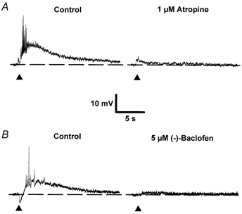
Figure 1. Effects of atropine and (-)-baclofen on the slow EPSPM.

A and B, single sweeps illustrating the effects of 1 μm atropine (A) and 5 μm (-)-baclofen (B) on slow EPSPs evoked in two separate neurones. The initial membrane potential of both cells was -64 mV. In B and in all subsequent figures, unless stated otherwise, traces are individual synaptic responses recorded intracellularly in response to a single stimulus delivered in the stratum oriens in the presence of 1-3 μm NBQX, 50 μm CGP 40116 and 50 μm picrotoxin. A, responses evoked in the additional presence of 1 μm CGP 55845A. Each sweep was taken at the same membrane potential achieved using DC injection to compensate for any drug-induced changes. Filled triangles mark the time of afferent stimulation. In all synaptic traces shown, action potentials are attenuated due to low sampling frequency.
