
Figure 6. Input to the apical tuft in the absence of background.
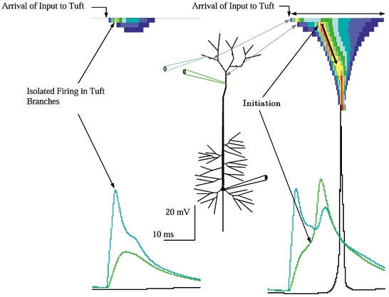
Left, just-subthreshold input. Just 2.5 nS per branch was adequate to trigger branch excitation, with the Na+-driven branch spikes reaching −15 mV. Note that despite the full blown event in the tuft branches firing was not triggered in the distal trunk, propagation inward to the soma did not ensue, and the tuft input was almost unobservable in the distal trunk. Right, threshold input to the apical tuft. With a small increase in input, to 4 nS per branch (20 nS total) firing spread from the sub-branches receiving input to abutting unstimulated sub-branches, similar to the spread within apical oblique branches, causing a bump in the declining phase of the voltage trace. The spread of excitation within the tuft and its closed end electrotonic geometry contributed to a rather sustained depolarization there, which in turn provided a sustained drive to the distal trunk, which after a delay for its charge-up reached its firing threshold at −43 mV (arrow). The delay between tuft and trunk indicated that excitation did not propagate from tuft into trunk, but rather that the tuft charged up the trunk over a period of 3–4 ms. The firing of the apex of the trunk was the critical event for firing the cell, because once initiated in the trunk propagation commenced inward, firing the soma when it arrived. Note the relatively abrupt upstroke of the action potential in the soma, a signature of the arrival of an inwardly propagating wave. The spatiotemporal plot reveals that the duration of depolarization in the tuft was quite sustained (two-headed arrow). This sustained tuft depolarization caused a sustained axial ohmic current flow into the distal trunk, which in turn drove voltage in the trunk section abutting the tuft to its Na+ threshold. Once initiated, the propagation inward proceeded (arrow with tail). Note the narrowing of the temporal (x-axis) width of the dendritic voltage wave as it propagated inward toward the soma.