
Figure 7. Tuft input triggers a distal Na+-Ca2+ spike which propagates inward.
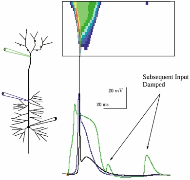
The response of a model neuron with increased dendritic Ca2+ conductance to synaptic input to apical tuft branches, in the absence of background. The Ca2+ current-driven width of the inwardly propagating voltage wave is greatest most distally and tapers as it travels inward, as can be seen in the spatiotemporal plot. Note that following the Ca2+ event subsequent input barrages were less effective in triggering firing. This suppression was due to dendritic [Ca2+]-gated K+ currents, and wore off with the same time course as Ca2+ recovery. As a result, for a 80-100 ms period following a distal Ca2+ event, repeated input was ineffective in driving the cell. Note that under these conditions (i.e. in the absence of background) the distal depolarization drives a pronounced after-depolarizing potential in the soma.