
Figure 9. Heparin does not interfere with the activation of ICRAC by passive store depletion or with the effects of 2-APB.
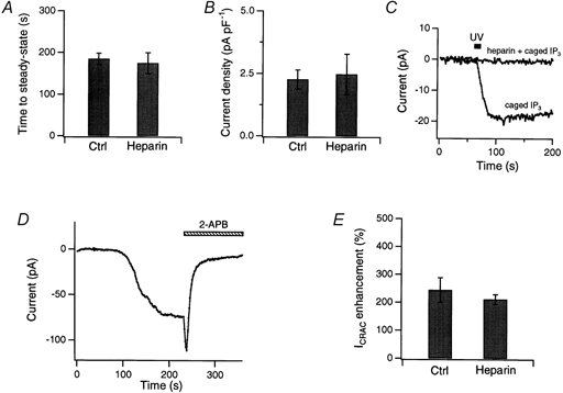
In all experiments except as noted, 500 μg ml−1 heparin was included in the recording pipette, and ICRAC was induced through passive depletion of internal stores by 10 mm EGTA in the patch pipette. A and B, heparin affects neither the rate of ICRAC activation (A) nor its maximal amplitude at steady state (B). The time to steady state was measured as the time to reach 95 % of the maximal current amplitude. Current amplitude was normalized to cell capacitance (average Cm= 9.95 pF). Heparin-treated cells (n = 6) are compared with matched control cells (n = 7). C, heparin loading via the patch pipette effectively inhibited IP3 receptors. In control Jurkat cells lacking heparin, ICRAC was activated by a 10 s UV flash to release caged IP3 (15 μm). With 500 μg ml−1 heparin in the pipette, uncaging IP3 failed to activate ICRAC. Results are representative of four cells loaded with caged IP3 alone and three cells with caged IP3 and heparin. D, 40 μm 2-APB first potentiated and then inhibited ICRAC in a Jurkat cell loaded with heparin via the patch pipette. Shown here are the peak currents during steps to −100 mV from +30 mV. E, summary of the potentiating effects of 5 μm 2-APB in heparin-treated (n = 6) and matched control (n = 5) cells.