
Figure 1. Membrane currents activated in rat portal vein myocytes by external application of ATP.
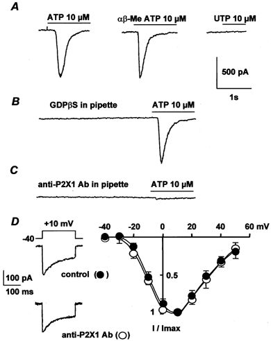
A, effects of 10 μm ATP, 10 μmαβ-MeATP and 100 μm UTP obtained from three different cells. B, pipette solution contained 2 mm GDPβS and the cell was dialysed with the pipette solution for 5 min before application of 10 μm ATP. C, intracellular application of 10 μg ml−1 anti-P2X1 antibody for 7 min before application of 10 μm ATP. In A–C, the myocytes were held at −60 mV. D, typical Ba2+ currents elicited by depolarization to 10 mV from a holding potential of −40 mV and current-voltage relationships obtained in control conditions (•) and after intracellular application of 10 μg ml−1 anti-P2X1 antibody for 7 min (○). Currents are expressed as a fraction of the maximal current (I/Imax) and are the means ±s.e.m. for 7–9 cells.