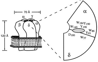
Figure 1.
Structural features of the nAChR. (Left) Global layout, adapted from data by Unwin (3), but with alternative ordering of subunits around the central axis (2, 20). (Right) Schematic of the agonist binding site (gray oval) viewed from the synapse, showing the large number of aromatic residues from three noncontiguous regions of the α subunit and key residues from the δ subunit also thought to contribute to binding. A comparable arrangement exists at the α/γ interface. Adapted from drawings previously presented by Galzi and Changeux (1) and Karlin and Akabas (2). Amino acid codes: W, tryptophan, Y, tyrosine, D, aspartate.

