
Figure 10. Diagrammatic representation of the involvement of 5-HT receptors in the control of the activity of BVPNs and CVPNs.
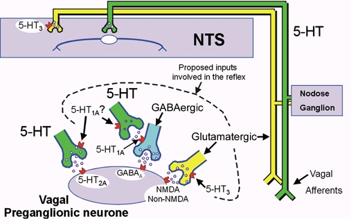
5-HT-containing neurones are shown in green, whereas glutamate-containing neurones are shown in yellow. These vagal preganglionic neurones can be activated reflexly via the nucleus tractus solitarii (NTS) by the cardiopulmonary afferents that run in the vagus nerve. It is proposed that the NTS neurones activate both a 5-HT-containing and a glutamatergic pathway. The 5-HT pathway inhibits the GABA-mediated (blue) ‘brake’, allowing the glutamatergic pathway to fully excite the preganglionic vagal neurones. This results in a bradycardia or bronchoconstriction. It should be noted that the 5-HT1A receptors are located presynaptically, not postsynaptically. In addition, the diagram shows a 5-HT-containing pathway directly innervating the vagal preganglionic neurones, which activates 5-HT2A receptors to cause excitation. This pathway is not believed to be involved in the reflex activation of vagal preganglionic neurones; however, it is speculated that the putative 5-HT1A receptors that mediate the inhibition of vagal preganglionic neuronal activity could be located on the nerve terminals of this pathway and, as such, would function as autoreceptors. It should be noted that these inhibitory 5-HT1A receptors could also be located on the terminals of the 5-HT-containing pathway, which inhibits the putative GABAergic ‘brake’. This figure and legend is a modification and update of that published by Ramage (2000).