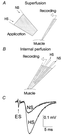
Figure 1. Local applications of HS to the synaptic area.

A, local superfusion of the synaptic area by HS or normal saline (NS) outside the recording pipette. The superfusion pipette was positioned within approximately 150–200 μm from the recording electrode. To avoid covering the synapse we used recording electrodes with a tip diameter of 3–5 μm. The tip diameter of the application pipette was 80 μm. B, solutions were changed inside the recording pipette of 10–20 μm tip diameter. C, the nerve extracellular spike (ES) was not affected by local applications of HS, while synaptic transmission was enhanced. Each trace represents the average of 600 EPSPs recorded at 1 Hz stimulation frequency.
