
Figure 1. PCR analysis of amplified aRNA from single respiratory brainstem neurons.
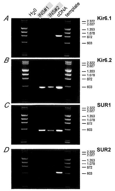
DNA fragments are amplified with primer pairs specific to Kir6.1 (A), Kir6.2 (B), SUR1 (C) and SUR2 (D). The first lanes next to the molecular weight marker (λHindIII-ΦXHaeIII digest) are H2O controls, templates in lanes 2 and 3 were of two different inspiratory neurons (INS#1, INS#2), and lane 4 used 0.1 ng of plasmid cDNA as positive control (except in D where 50 ng of atrial cDNA was used). Primers were sensitivity-tested to amplify fragments of 865 bp (Kir6.1), 553 bp (Kir6.2), 539 bp (SUR1) and 603 bp (SUR2). Fragment sizes of the molecular marker are indicated on the right. Note that fragments amplified from single respiratory neurons can only be detected for Kir6.2 and SUR1.