
Figure 1. Diagram showing functional organization of ICC in the canine gastric antrum.
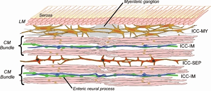
The antral wall contains two layers of smooth muscle cells: the outer longitudinal muscle layer (LM) lacking ICC and the inner circular muscle layer (CM) in which individual smooth muscle cells are organized into bundles. In this layer ICC-IM are distributed through individual CM bundles. A network of ICC lies between the LM and CM (ICC-MY). In CM bundles, ICC-IM function both to augment depolarizations reaching them from ICC-MY and as essential intermediaries in the transmission of information from enteric neural processes to nearby smooth muscle cells. ICC-SEP form functional cables which transmit information from pacemaker ICC-MY to deeper CM bundles present in the stomachs of larger animals. Although not demonstrated as yet, it may well be that ICC-SEP and ICC-MY are directly connected; alternatively ICC-SEP may be electrically excited by activity in CM bundles lying closer to the ICC-MY pacemaker network.