
Figure 1. Examples of EPSPs (*) and IPSPs (†) in recordings from two types of PO/AH neurones.
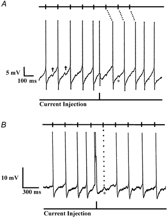
All action potentials have been truncated. Halfway through each record, a 2 ms 200 pA depolarizing current pulse produced a premature action potential. Tick marks at the top of each record show the predicted times of action potentials, based on the firing pattern before the current pulse. A, spontaneously firing, warm-sensitive neurone displaying slow, depolarizing prepotentials preceding each action potential. After the premature action potential at the current pulse, all subsequent action potentials were shifted in time, suggesting that the firing pattern was intrinsically generated. B, EPSP-driven neurone whose firing pattern was not altered by the current injection. Note the EPSPs prior to each action potential and the prominent EPSP without an action potential at the first tick mark (dotted line) after the current pulse.