
Figure 6. Morphological and physiological characteristics of a temperature-insensitive preoptic neurone recorded in a coronal tissue slice.
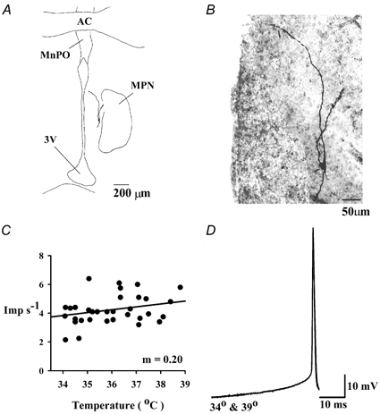
A, camera lucida drawing of the biocytin-filled neurone. B, photograph of the biocytin-filled neurone. Dorsal is at the top. 3V, third ventricle; MPN, medial preoptic nucleus; MnPO, median preoptic nucleus; AC, anterior commissure. The left side of the photograph (B) is the edge of the third ventricle. C, effect of temperature on the neurone's firing rate (imp s−1). The regression coefficient, m, defines the neurone's thermosensitivity, which was 0.20 imp s−1°C−1. Each point represents a sample taken every 10 s. D, superimposed averages of 15 action potentials recorded at two different temperatures, 34 and 39 °C. Warming to 39 °C had no effect on the depolarizing prepotential.