
Figure 8. Camera lucida drawings showing projected locations and morphologies of temperature-insensitive neurones (left side) and warm-sensitive neurones (right side) recorded in horizontal tissue slices.
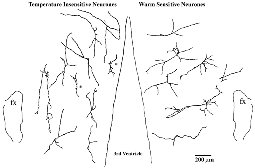
Neurones identified at different dorsal-ventral locations are projected onto a single horizontal plane. Rostral is at the top; fx, fornix. Cells marked with asterisks had dendrites shorter than 200 μm and were not included in the averages shown in Fig. 9 (see Methods).