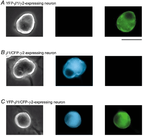
Figure 1. Heterologous expression of YFP-β1 and CFP-γ2 alone or in combination in adult rat SCG neurons.

Phase contrast and fluorescence images of neurons expressing YFP-β1/γ2 (A), β1/CFP-γ2 (B) and YFP-β1/CFP-γ2 (C). Fluorescence images were taken with a filter set specific for CFP (centre panels) and YFP (right panels; 440 nm excitation, and 480 nm emission and 535 nm emission for CFP and YFP, respectively). Fluorescence images shown are pseudocoloured. Scale bar, 55 μm.
