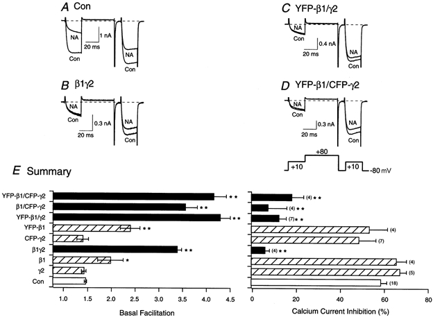
Figure 2. Effect of heterologous expression of β1, YFP-β1, γ2 or CFP-γ2 alone or in combination on facilitation and noradrenaline (NA)-mediated inhibition of ICa in SCG neurons.

Superimposed ICa traces evoked with the double-pulse voltage protocol (shown at the bottom of D) in the absence (lower traces) and presence (upper traces) of 10 μm NA for control (A), β1γ2-expressing neurons (B), YFP-β1/γ2-expressing neurons (C) and YFP-β1/CFP-γ2-expressing neurons (D). Currents were evoked every 10 s. Dashed lines indicate the zero current level. E, summary graphs of mean (±s.e.m.) basal facilitation and ICa inhibition for neurons expressing β1, YFP-β1, γ2 or CFP-γ2 alone or in combination. The final concentration of cDNA injected was 10 ng ml−1 per subunit. Facilitation was calculated as the ratio of ICa amplitude determined from the test pulse (+10 mV) occurring after (postpulse) and before (prepulse) a +80 mV conditioning pulse. *P < 0.05 and **P < 0.01 vs. control (uninjected neurons, Con). Numbers in parentheses indicate the number of experiments.
