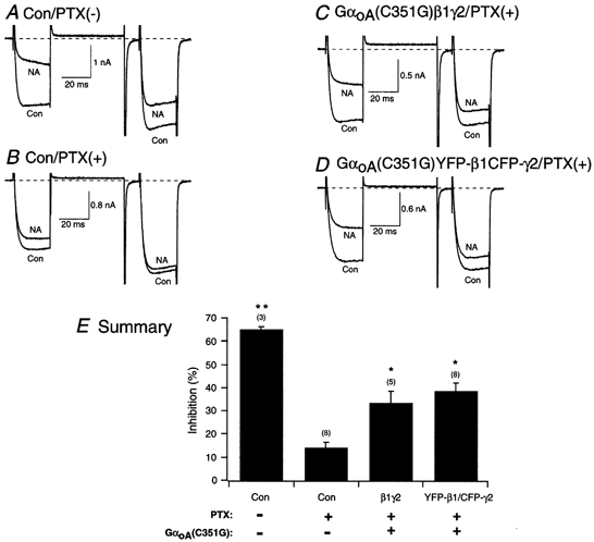
Figure 5. Reconstitution of α2-adrenergic receptor coupling to N-type Ca2+ channels in SCG neurons expressing PTX-resistant GαoA with β1γ or YFP-β1/CFP-γ2.

Superimposed ICa traces evoked with the double-pulse voltage protocol (shown at the bottom of Fig. 2D) in the absence (lower traces) and presence (upper traces) of 10 μm NA for control, no PTX (A), control plus PTX (500 ng ml−1, overnight; B), PTX-pretreated neuron expressing GαoA(C351G; C) and PTX-pretreated neuron co-expressing GαoA(C351G) and YFP-β1/CFP-γ2 (D). Currents were evoked every 10 s. Dashed lines indicate the zero current level. E, summary graph of mean (±s.e.m.) NA-mediated ICa inhibition for uninjected (Con), GαoA(C351G)/β1/γ2-expressing neurons and GαoA(C351G)/YFP-β1/CFP-γ2-expressing neurons. NA-mediated inhibition was determined as described in the legend of Fig. 2. The final concentration of cDNA injected was 10 ng ml−1 for the β and γ subunit, and 7 ng ml−1 for the PTX-resistant α subunit. *P < 0.05 and **P < 0.01 vs. PTX-treated control neurons. Numbers in parentheses indicate the number of experiments.
