
Figure 3.
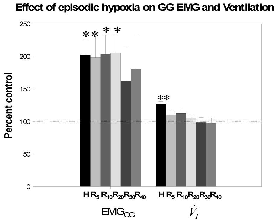
Group data (all subjects) for phasic EMGGG and V̇I during hypoxia and recovery periods normalized to baseline as a percent of control. H represents averaged data of the hypoxia trials while R5, R10, R20, R30, R40 are averaged data as a percent of room air control during recovery 5, 10, 20, 30 and 40 minutes, respectively. Both V̇I and phasic EMGGG activity increased significantly during hypoxia vs room air control. However, only EMGGG phasic activity was significantly increased during recovery periods R5, R10, and R20, as a percent of room air control; **p<0.05, hypoxia vs control period; *p<0.01, hypoxia, R5, R10, R20 vs control period. While phasic EMGGG remained elevated in R30 and R40, the values did not reach statistical significance due to the small number of subjects with stable sleep during this period, see text for details.