
Figure 4.
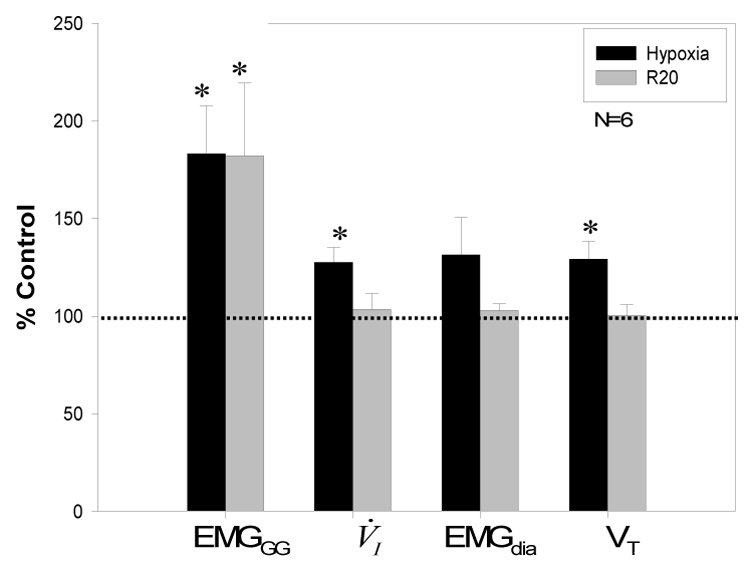
Group data for 6 subjects with satisfactory EMGdia signals are presented. Data for phasic EMGGG, V̇I, EMGdia and VT during hypoxia and recovery period (R20) were normalized to baseline as a percent of control. Phasic EMGGG, V̇I, and Vt, all increased significantly during hypoxia vs room air control. However, only phasic EMGGG activity was significantly increased during R20; *p<0.05.