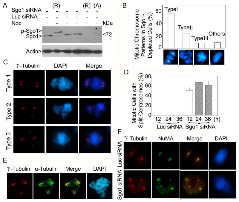
Figure 1.
siRNA depletion of Sgo1 causes formation of extra γ-tubulin foci. (A) HeLa cells were transfected with Sgo1 or luciferase (Luc) siRNA for 24 h. Rounded-up (R) and adherent (A) cells in Sgo1 siRNA transfected dishes were collected separately. An equal amount of each cell lysate was blotted for Sgo1 and β-actin. Nocodazole (Noc)-treated cell lysates were loaded as control. Arrow p-Sgo1 indicates phospho-Sgo1. (B) Rounded-up cells induced as a result of Sgo1 depletion were examined for chromosome patterns after DAPI staining. The data were summarized from more than 300 mitotic cells depleted of Sgo1. The stars (*) denote spindle pole positions. (C) HeLa cells transfected with Sgo1 siRNA were stained with an antibody to γ-tubulin (red). DNA was stained with DAPI (blue). Representative images of cells with centrosome splitting are shown. (D) The percentage of siRNA-transfected cells with extra centrosomal foci was summarized from 200 mitotic cells at each time point. (E) A representative cell transfected with Sgo1 siRNA for 24 h and stained with antibodies to γ-tubulin (red) or α-tubulin (green) is shown. (F) HeLa cells transfected with Sgo1 or Luc siRNA were stained with antibodies to γ-tubulin (red) and NuMA (green). Representative images are shown.

