
Figure 6.
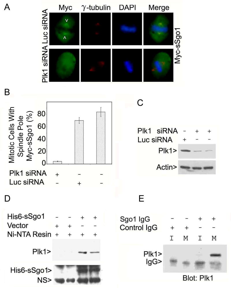
Plk1 is physically associated with sSgo1. (A) HeLa cells co-transfected with Myc-sSgo1 plasmid and Plk1 or Luc siRNA for 24 h were stained with antibodies to the Myc tag (green) and γ-tubulin (red). (B) HeLa cells were cotransfected with Myc-sSgo1 expression plasmid and Plk1 or Luc siRNA. The data were summarized from three independent experiments (100 Plk1-depleted mitotic cells/experiment). (C) HeLa cells transfected with Plk1 or Luc siRNA for 24 h were blotted for Plk1 and β-actin. (D) Aqual amounts of HeLa cell lysates after transfection with a His6-sSgo1plasmid or the vector alone for 48 h were incubated with Ni-NTA resin. Proteins specifically bound to the resin were blotted for Plk1 or Sgo1. Arrow NS denotes a non-specific signal. (E) An equal amount of interphase (I) or mitotic (M) HeLa cell lysate was immunoprecipitated with the anti-Sgo1 antibody or with a control IgG. The immunoprecipitates were blotted for Plk1.