
Fig. 1.
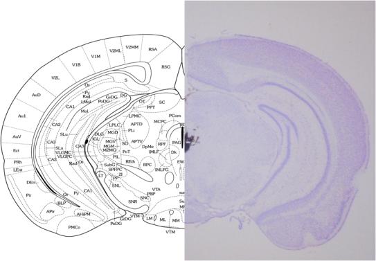
Photomicrograph of cresyl violet-stained coronal mouse brain section and corresponding atlas plate (Franklin and Paxinos, 2001) indicating the typical site and spread of microinjection as assessed by stain injections. All doses of β-CNA and TRIM were injected at a volume of 0.5 ml into the PAG.