
Figure 5.
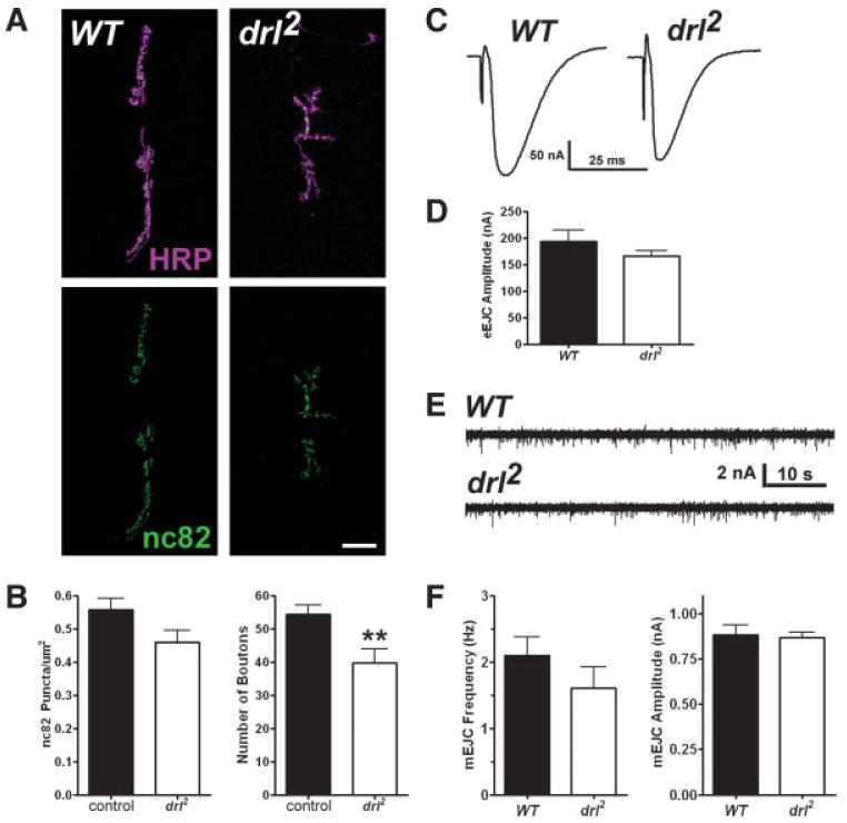
drl mutant animals exhibit a reduction in the number of boutons but not active zones. (A) Confocal fluorescent images showing NMJs labeled with antibodies against HRP (magenta) and the presynaptic protein Brp (nc82, green). Scale bar: 20 μm. (B) Quantification of active zone density (nc82 puncta/μm2, left) and bouton numbers (per NMJ, right). (C) Representative two-electrode voltage clamp recordings from muscle 6 of control and drl2 third instar larvae, showing evoked endplate junctional currents (EJCs). (D) Quantification of EJC amplitudes. (E) Representative voltage clamp recordings from control and drl2 third instar larvae, showing spontaneous excitatory junctional currents (mEJCs) in the muscle 6 NMJ. (F) Quantification of mEJC frequency (left) and amplitude (right). [Color figure can be viewed in the online issue, which is available at www.interscience.wiley.com.]