Figure 5.
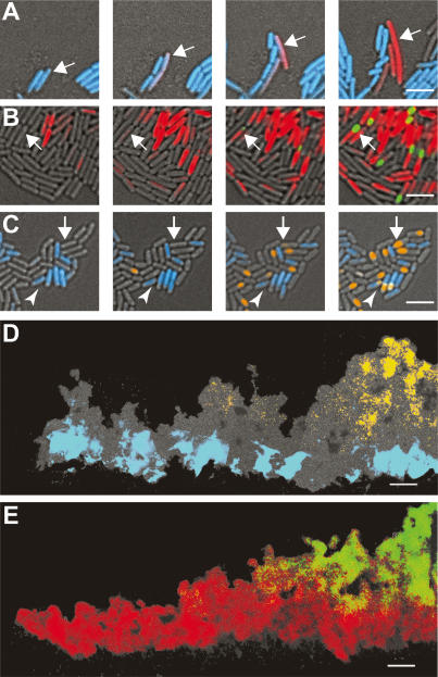
Developmental history of cells within a biofilm. Time-lapse images of cells harboring dual reporter fusions. (A) Images of cells harboring Phag-cfp (motile cells, blue) and PyqxM-yfp (matrix-producing cells, red) were taken every 1 h. Arrow indicates a motile cell transitioning to matrix-producing cell. (B) Images of cells harboring PyqxM-cfp (matrix-producing cells, red) and PsspB-yfp (sporulating cells, green) were taken every 2 h. Arrow highlights a cell initiating matrix production and then transitioning to a sporulating cell. (C) Images of cells harboring Phag-cfp (motile cells, blue) and PsspB-yfp (sporulating cells, orange) were taken every 2 h. The majority of the sporulating cells arise from nonmotile cells, indicated by the white arrow. The arrowhead highlights an example of the minority of motile cells that initiate sporulation. (D,E) Thin sections of 48-h colonies from cells harboring dual reporters. Images formatted as in Figure 3. In D, motile cells (blue) appear in distinct regions relative to sporulating cells (orange), whereas in E, matrix-producing cells (red) overlap with the region of sporulating cells (green). Bars: A–C, 5 μm; D,E, 50 μm.

