
Figure 3.
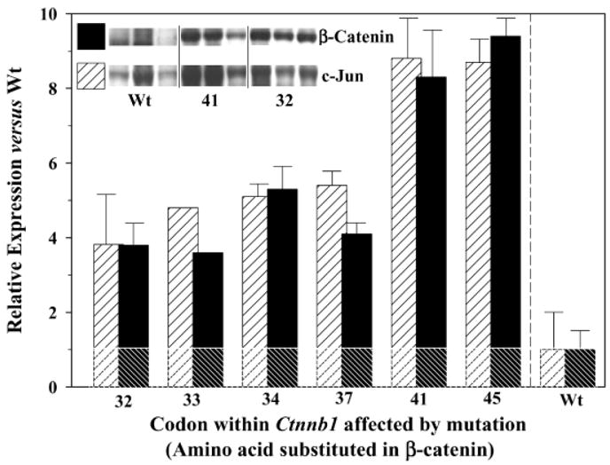
Expression in rat colon tumors of β-catenin (filled bars) and c-jun (hatched bars). Total tissue lysates were prepared from three separate colon tumors for each of the codons mutated in Ctnnb1, and these were subjected to SDS-polyacrylamide gel electrophoresis and probed with antibodies to β-catenin and c-Jun proteins, as reported before [16]. Inset, representative blots from three tumors containing wild-type (wt) β-catenin, three tumors with mutations in codon 41 of Ctnnb1, and three tumors with mutations in codon 32 of Ctnnb1. Normal colonic mucosa and positive controls for the proteins of interest were included in each gel (not shown). Densitometry measurements were obtained with a gel documentation system and associated software; results were first normalized to expression levels of glyceraldehyde-3-phosphate dehydrogenase and then expressed relative to the protein levels in tumors with wild-type β-catenin (assigned an arbitrary expression value of 1.0). Data represent mean ± SD, n = 3 (except for codon 33, which was from a single tumor). The horizontal line is a reference guide indicating the relative expression of 1.0 for c-jun and β-catenin proteins in tumors with wt β-catenin; all of the bars above this line were significantly different from wt controls, and the bars labeled 41 and 45 were significantly different from all others (P < 0.05).