
Fig. 1.
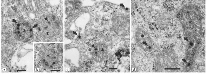
Electron micrographs of bovine adrenal chromaffin cells (a-c) and mouse pheochromocytoma cells (d) infected with a recombinant non-replicating adenoviral vector. a, b, d Mitochondria at various stages of degeneration (asterisks) with deteriorated internal membrane and several adenoviral particles inside. c A mitochondrion with healthy internal structure and remnants of viral particles inside. Arrows = intact viral particles; arrowheads = viral particles at various stages of breakdown. Bar = 500 nm.