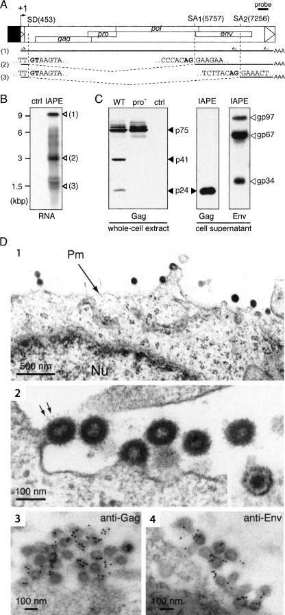
Figure 3.
Structure of the IAPE-D1 provirus and characterization of its gene products. (A) Genomic organization of the IAPE-D1 provirus, cloned under the control of the CMV promoter (in black) and structure of the corresponding viral transcripts. (SD) Splice donor site; (SA) splice acceptor site. The splice sites of the IAPE subgenomic transcripts (2 and 3) were determined by sequencing the RT-PCR products obtained using total RNAs and the two sets of primers indicated. (B) Northern blot analysis of total RNA extracted from 293T cells transfected with pCMV-β (ctrl) or the IAPE-D1 expression vector (IAPE), using a probe (shown in A) complementary to a 3′ domain of the IAPE genome. (C) Western blot analysis of IAPE-D1 proteins. Whole-cell extracts and virion-containing supernatants of human 293T cells transfected with IAPE-D1 (WT), a protease-deficient mutant (pro−), or a control plasmid (ctrl) were analyzed by Western blotting, using rabbit anti-IAPE Gag or anti-IAPE Env antisera, as indicated. (D, 1–4) Electron microscopy of cells transfected with IAPE-D1 and of the released viral particles. (1) Representative low-magnification image of transfected 293T cells, with particles budding at the plasma membrane. The nucleus (Nu) and plasma membrane (Pm) are indicated. (2) High-magnification view of budding and extracellular particles. Prominent spikes, corresponding to the Env protein, are indicated (arrows); (inset) view of an extracellular mature particle, with a condensed central core. (3–4) Immunogold labeling of IAPE particles in transfected 293T cells using the anti-IAPE Gag (3) or anti-IAPE Env (4) rabbit antiserum and a secondary antibody linked to gold beads, observed by electron microscopy. Gold beads are preferentially associated with viral particles (439 ± 165 gold beads/μm2 associated with VLPs vs. 5.6 ± 3.0 and 2.8 ± 2.6 gold beads/μm2 associated with cytoplasm and VLP-free extracellular medium for the anti-IAPE Gag antiserum and 302 ± 119 gold beads/μm2 associated with VLPs vs. 18.1 ± 9.1 and 4.2 ± 2.4 gold beads/μm2 associated with cytoplasm and VLP-free extracellular medium for the anti-IAPE Env antiserum).

