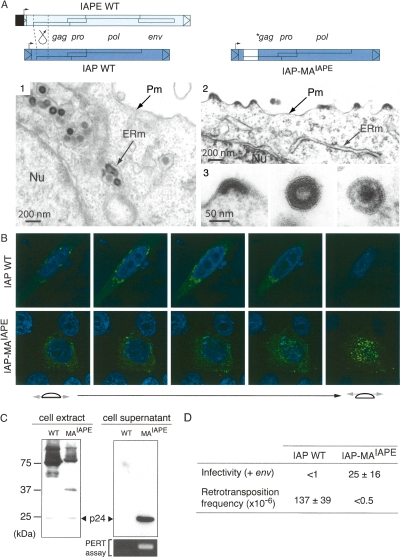
Figure 5.
Recovery of an extracellular life cycle for an IAP chimera. (A) Structure of the wild-type and chimeric IAPs and morphology of associated VLPs. The IAP-MAIAPE element was constructed as illustrated by replacing the IAP Gag N terminus with that of IAPE. (1–3) Electron microscopy of 293T cells transfected with either wild-type or chimeric IAPs. (1) Representative low-magnification image of wild-type IAP particles accumulated in the cisternae of the ER. No particle can be observed at the cell membrane or in the extracellular space. (2) Low-magnification image of cells transfected with the chimeric IAP, disclosing particles budding at the cell membrane. No particle can be observed in the ER. (3) High-magnification images of chimeric IAP-associated particles. Images represent (from left to right) budding, free immature, and free mature particles. (B) Immunofluorescence confocal analysis of human HeLa cells transfected with wild-type or chimeric IAPs, fixed, permeabilized, and stained with the anti-Gag antibody (in green). Nuclei are stained with TO-PRO-3 iodide (in blue). (C) Western Blot analysis of whole-cell lysates or cell supernatants from 293T cells transfected with either the wild-type (WT) or the chimeric IAP (MAIAPE). (Bottom right) Detection of RT activity in the corresponding cell supernatant, using the PERT assay. (D) Functional characterization of wild-type and chimeric IAPs. For the intracellular retrotransposition assay, HeLa cells were cotransfected with the wild-type or chimeric IAP, and a neoTNF-marked defective IAP reporter in which a (backward) neomycin resistance gene—with the neo ORF interrupted by a (forward) intron—becomes functional only after splicing out of the intron, upon achievement of a complete replicative cycle. Retrotransposition events are detected upon G418 selection of the transfected cells, and frequencies are expressed as numbers of G418R clones per transfected cells in selection (mean values ± SD, n = 4); see Dewannieux et al. (2004) and Supplemental Figure S2 for a detailed description of the assay. For the infectivity assay, 293T cells were cotransfected with the same IAP plasmids as above, but with in addition the IAPE-D1 Env expression vector. Supernatants from the transfected cells were collected and used to infect naive HeLa cells, as in Figure 4A, and the viral titers were determined upon G418 selection of the target cells (mean values ± SD, n = 3).

