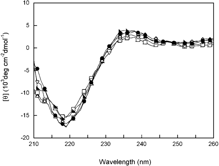
Figure 2.
Far-UV CD of wild-type (◂), M43A (□), F56A (▾), I81A (▸), V132A (♦), L145A (▵) and V170A (•) HγD-Crys. Samples contained 100 μg/mL protein in 10 mM sodium phosphate, 5 mM DTT, 1 mM EDTA (pH 7.0) at 37°C. A 0.25-cm pathlength cuvette was used for all measurements. All spectra were corrected for background buffer signal.

