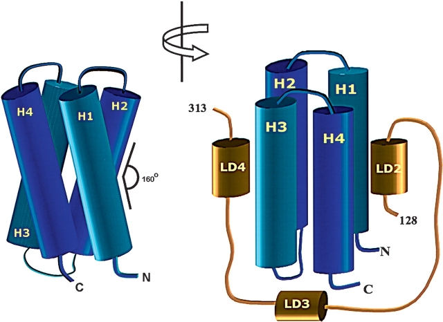
Figure 7.
A model of paxillin binding to the FAT domain of FAK. The FAT domain is an elongated four-helix bundle with a right-hand twist (A). The LD2 and LD4 motifs of paxillin bind to opposite faces of the four-helix bundle of FAT but are oriented in the same direction (B). The intermediate residues of paxillin remain unstructured in the complex and probably wrap around the H3–H4 side of FAT, although no further binding interactions are observed.

