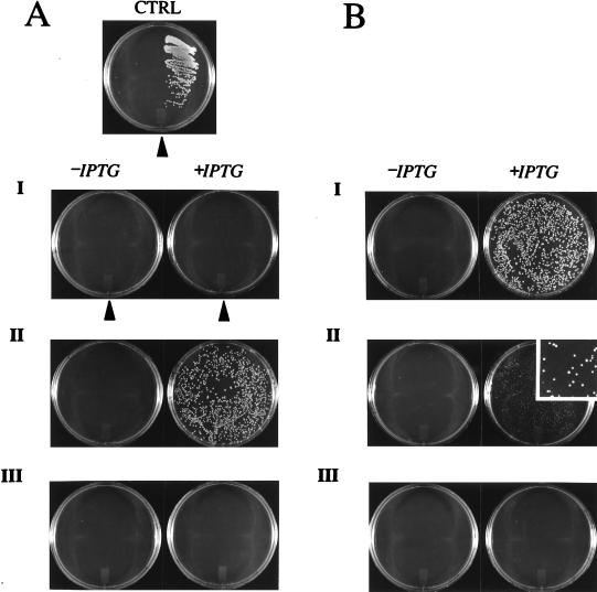
Figure 3.
(A) E. coli survival assay on minimal medium plates. Control, left side of the plate: E. coli harboring pQE-30 (no insert); right side: E. coli expressing full-length mDHFR. (A, I) Left half of each plate: transformation with Z-F[1,2]; right half of each plate: transformation with Z-F[3]. (A, II) Cotransformation with Z-F[1,2] and Z-F[3]. (A, III) Cotransformation with constructs control-F[1,2] and Z-F[3]. All plates contain 0.5 μg/ml trimethoprim and plates on the right side of I-III contain 1 mM IPTG. (B) E. coli survival assay using destabilizing DHFR mutants. (B, I) Cotransformation with Z-F[1,2] and Z-F[3:Ile-114 → Val]. (B, II) Cotransformation with Z-F[1,2] and Z-F[3:Ile-114 → Ala]. (Inset) A 5-fold enlargement of the right-side plate. (B, III) Cotransformation with Z-F[1,2] and Z-F[3:Ile-114 → Gly]. All plates contain 0.5 μg/ml trimethoprim and plates on the right side contain 1 mM IPTG.

