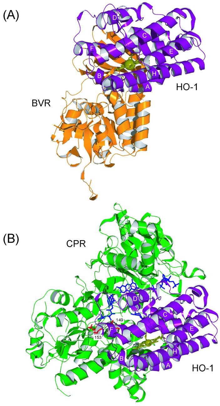
Fig. 3. Putative docking model of rat HO-1 (PDB 1DVE) with rat BVR (1GCU) (A) and with CPR (1AMO) (B; ref 16) calculated by the program Hex.
HO-1 is represented by a ribbon (purple). Heme is shown as ball-and-sticks (olive). The side chains of Lys-149 and Lys-153 in F helix of HO-1 are represented in stick form (red). BVR and CPR are shown as orange and green ribbons, respectively. The cofactors of CPR are in stick form (blue).

