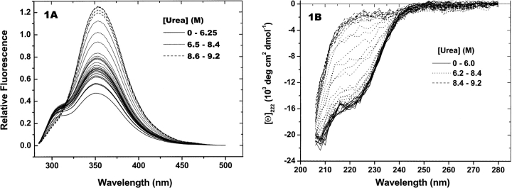
Figure 1.
rhm CSF-β titration spectra. rhm CSF-β (0.3 mg/mL) was titrated with urea, as described in Materials and Methods. Heavy lines are spectra collected at the urea concentrations shown in the legends. The thin solid lines are spectra identified as pretransition and posttransition spectra. Thin dotted lines are spectra measured over the structural transition. (A) Fluorescence spectra. (B) CD spectra.

