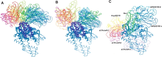
Figure 5.
Initial (A) and final (B) structures of the substrate binding domain (SBD) docked with the ATPase/J-domain complex. (C) The DOT and ZDOCK docking sites for the SBD. The ATPase domain is shown in blue, and in A and B the J-domain is in magenta. In C, the NoJ docking site for the SBD occupies the J-domain binding site. In the final structures (B) the clump of structures to the upper right of the J-domain corresponds to the HPD-loop/D152 set, and the clump to the upper left corresponds to the positive-patch/E218 set.

