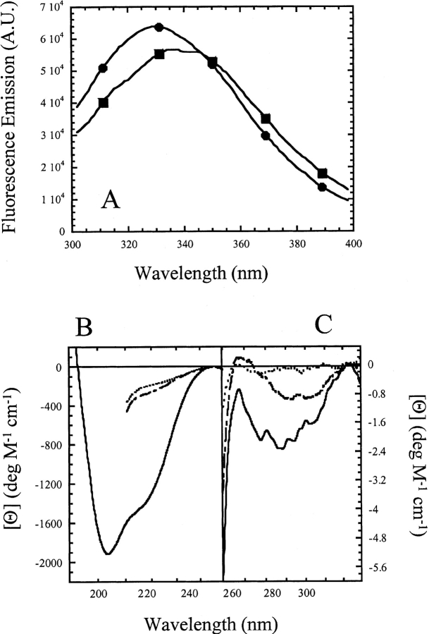
Figure 2.
(A) Fluorescence emission spectra of CP1-CARD. The protein (3 μM) in buffer (circles) or in 5.8 M urea-containing buffer (squares) was excited at 280 nm. The fluorescence emission was collected from 300 to 400 nm. (B) Far-UV and (C) near-UV CD spectra of CP1-CARD. The spectra of native protein in buffer (solid line) and protein unfolded in 6 M urea-containing buffer (long dashed line) or 6 M guanidine-containing buffer (short dashed line) are shown.

