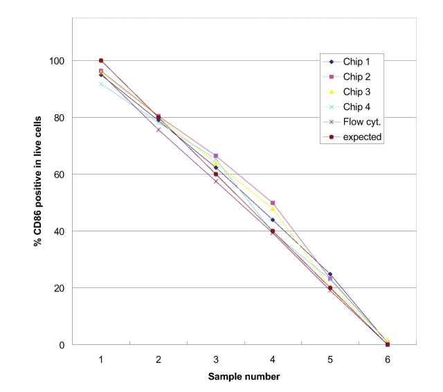
FIGURE 3.
Comparison between on-chip CD86 antibody staining results from four different experiments. The data were obtained with classically stained samples measured on a conventional flow cytometer. Data are in good agreement with those obtained by the reference technique and the theoretical prediction.

