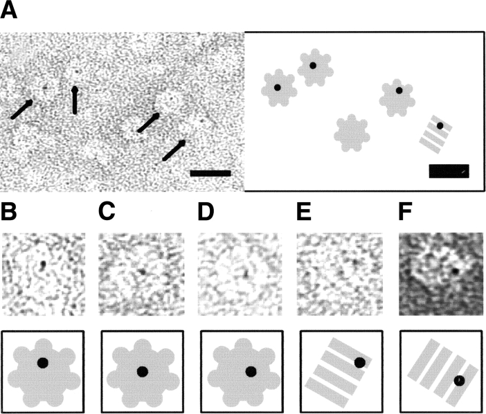
Figure 6.
Electron micrographs of negatively stained S105-nanogold conjugates in the presence of GroEL. GroEL molecules labeled with S105-Nanogold can be clearly discerned, and are highlighted by arrows in the overview (A) and individually boxed molecules (B–F). Note that (A–E) were stained with methylamine vanadate (Nanovan); (F), with ammonium molybdate. The scale bar corresponds to 20 nm. A cartoon interpretation of each panel is provided to highlight top-down and side-on GroEL molecules.

