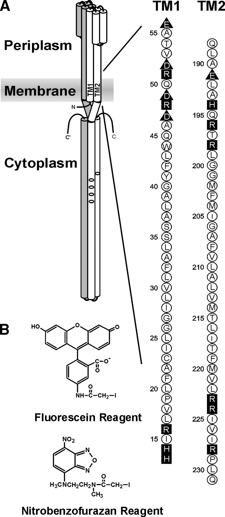
Figure 1.

Chemoreceptor organization, the transmembrane regions of Trg, and the fluorescent sulfhydryl reagents. (A) (Left) A cartoon of the organization of a chemoreceptor homodimer. Amino (N) and carboxyl termini (C) are labeled, and one subunit is shaded. On the other subunit, the transmembrane regions TM1 and TM2 are labeled and the positions of methyl-accepting sites shown by ovals. The approximate position of the cytoplasmic membrane is shown as a shaded area. (Right) The amino acid sequences (single letter code) of the TM1 and TM2 regions of Trg. Neutral and hydrophobic residues are enclosed in open circles; charged residues, in dark symbols; negative charges, in triangles; and positive charges, in squares. (B) Structure of 5-IAF (Fluorescein Reagent) and N, N′-dimethyl-N-(iodoacetyl)-N′-(7-nitrobenz-2-oxa-1, 3-diazol-4-yl)ethyl-ene-diamine (Nitrobenzofurazan Reagent).
