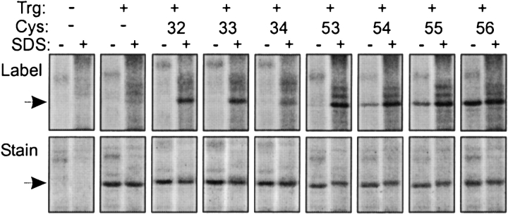
Figure 2.
Labeling of cysteine-containing forms of Trg by a fluorescent sulfhydryl reagent. Trg lacking cysteine or containing a single cysteine at the indicated positions in the TM1 region was treated with 5-IAF in its native, membrane-embedded state (−) or in the presence of the denaturant and membrane solvent SDS (+), and separated from other proteins in the membrane vesicles by SDS-PAGE. The figure shows two fluorographic images of the same segments of SDS polyacrylamide gels, the upper ones generated by fluorescein fluorescence, and the lower ones, by the protein stain SYPRO red. The leftmost pairs of lanes show patterns for vesicles lacking Trg. The position of Trg, Mr ~60,000, is shown by the arrows.

