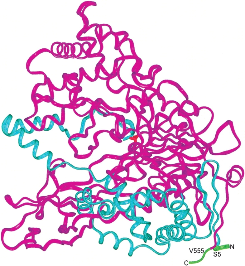
Figure 1.

The PGA dimeric structure. The β-subunit is shown in magenta, and the α-subunit, in blue ribbons. The polypeptide regions trimmed from the N terminus of the α-subunit and from the C terminus of the β-subunit are indicated in green. The amino acid residues to be connected with the four amino acids linker are labeled. In red, at the center of the molecule, the catalytic serine residue is indicated.
