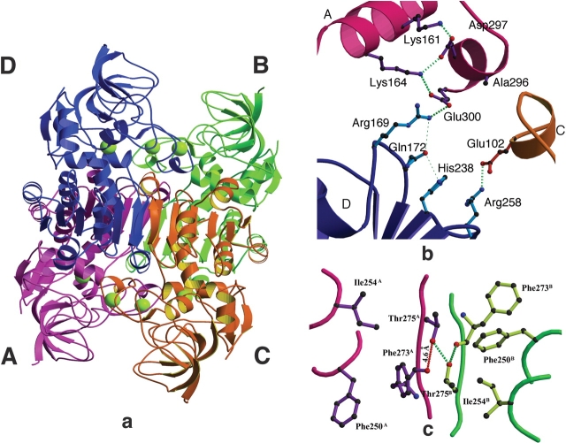
Figure 4.
(a) The tetramer of PaADH. Subunits A, B, C, and D are colored purple, green, orange, and blue, respectively. The catalytic and structural Zn2+ ions are shown in yellow. (b) The ion-pair network at the interface of three subunits of PaADH. The long ion-pair network consists of Lys 161A-Asp 297A-Lys 164A-Glu 300A and Arg 169D (superscripts denote the different subunits). The short ion pair includes Arg 258D and Glu 102C. The ion pairs are shown as thick dotted green lines. Subunit assignment and coloring are as in the legend to Figure 4a, above. (c) The interface of subunits A and B. At the dimer interface, a distortion of the noncrystallographic twofold symmetry is due to the asymmetrical, donor–acceptor nature of the intersubunit Thr 275A–Thr 275B hydrogen bond. The hydrogen bonds are shown in green dashed lines; the 4.6 Å distance between the Thr 275A hydroxyl and the Phe 273A carbonyl is shown in red (dashed line).

