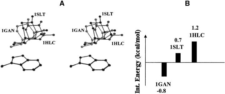
Figure 8.
(A) Stereo diagram showing superposition of the binding site aromatic residues along with the bound galactose in toad ovary galectin (1GAN), S-lac lectin (1HLC), and S-lectin (1SLT). Superposition has been carried out with reference to the atoms of the aromatic residue. The position-orientation of the bound saccharide with respect to the aromatic residue is similar in these three proteins of the galectin family. The main chain atoms of aromatic residues are not shown for clarity. (B) Bar diagram showing the interaction energies of galactose/3-methylindole complexes corresponding to the position-orientation observed in these three proteins.

