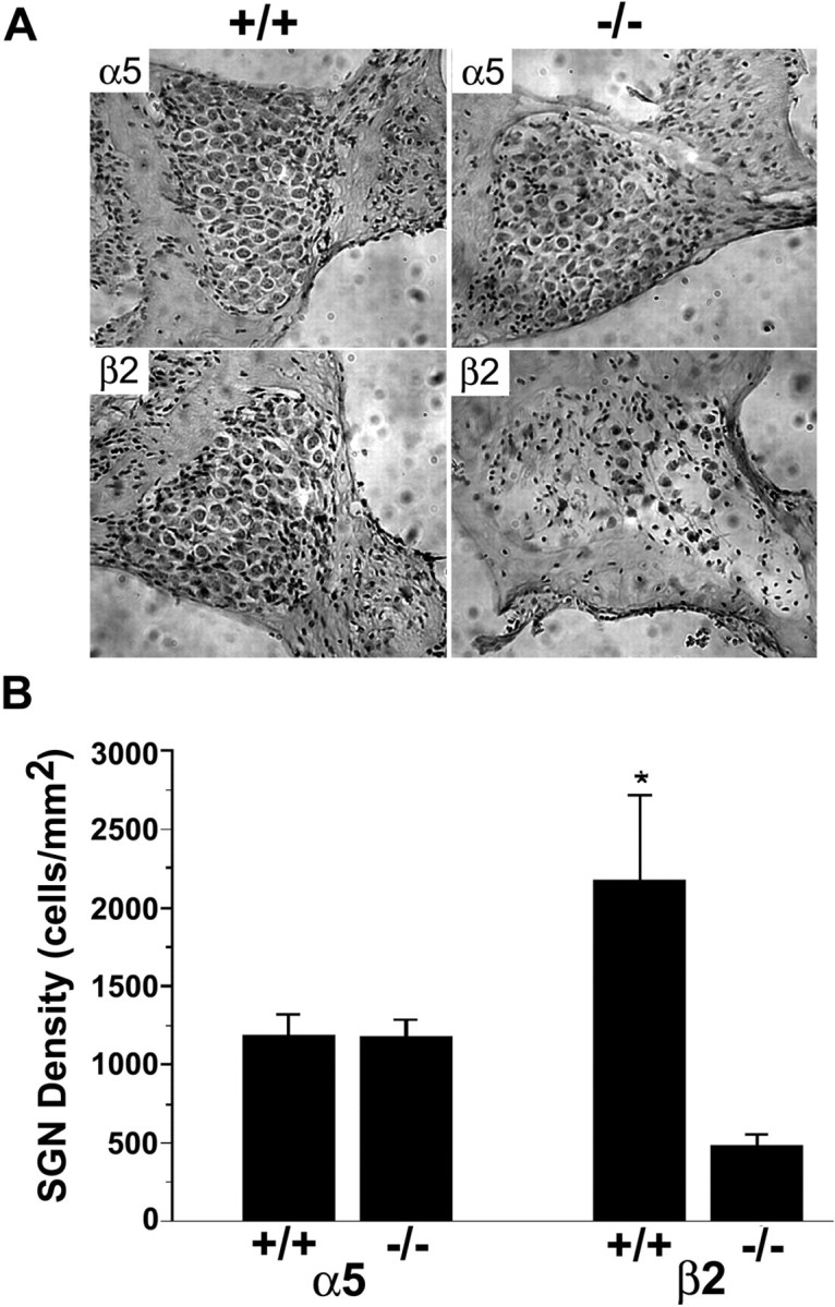
Figure 3.

Histology of spiral ganglia from α5 or β2 null mice. The top shows mid-modiolar sections of spiral ganglion from one 8-month-old mouse lacking α5 (right) and one 8-month-old α5+/+ control mouse (left) with the same genetic background (A). The bottom consists of mid-modiolar sections of spiral ganglion from one 8-month-old mouse lacking β2 (right) and one 8-month-old β2+/+ control mouse (left) with the same genetic background. Dramatic loss of SGNs can be observed in the section of spiral ganglia from 8-month-old mice lacking the β2 subunit (A). In B, the right basal area of the spiral ganglion and the number of SGNs were quantified in every other section using Image Pro Plus (Media Cybernetics). Each group contains three 8-month-old mice. The density of SGNs in β2-/- mice was significantly decreased compared with genetic-matched controls (*p < 0.01). Error bars represent SEM.
