Figure 4.
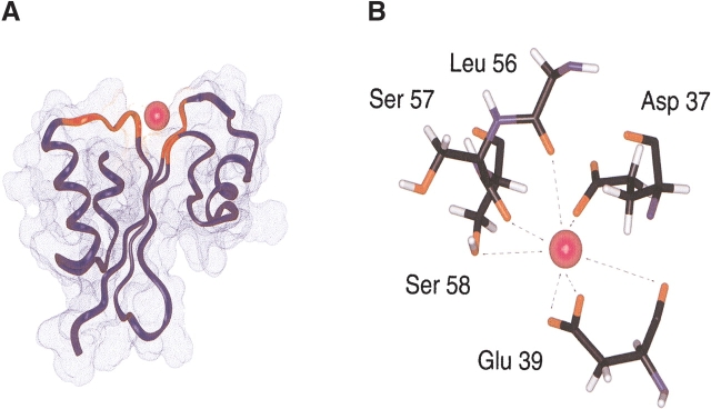
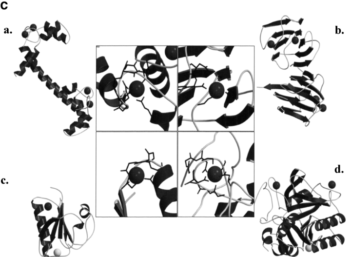
Calcium binding motif of MTH1880. (A) The structural model of MTH1880 bound to a calcium ion. Regions in red color represent the residues whose resonances changed upon calcium binding. (B) Proposed coordination of the calcium binding site based on NMR data. The residues involved in calcium binding were displayed with side-chain atoms. (C) A class of calcium binding motifs. (a) Calmodulin (PDB: 3CLN), (b) crystallin (PDB: 1HDF), (c) gelsolin (PDB: 4CPV), and (d) thermitase (PDB: 1THM). The calcium ion is shown as a black sphere and sodium ion as a white sphere.


