Figure 8.

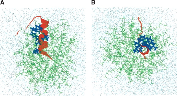
Initial conformation of hydrated FP-SDS micellar system before molecular dynamics simulations, viewed from the side (A) and down (B) the peptide α-helix (FP residues 3–16). (A) Side view of the initial configuration of the hydrated FP-SDS micelle, with the peptide’s α-helix (residues 3–16) penetrating the micelle, whereas the hydrophilic C-terminal region (residues 17–23) wraps around the micellar surface (i.e., micelle–water interface). The SDS detergent lipids are represented by green stick models; the FP backbone, as a red ribbon with the side chains for Ile-4, Leu-7, Phe-8, Leu-9, Phe-11, and Leu-12 as blue stick models; and the water molecules and sodium and chloride ions by dots. (B) Top view of the same initial configuration for the FP-SDS micellar system. The overall initial configuration of the FP-SDS micelle is spherical.
